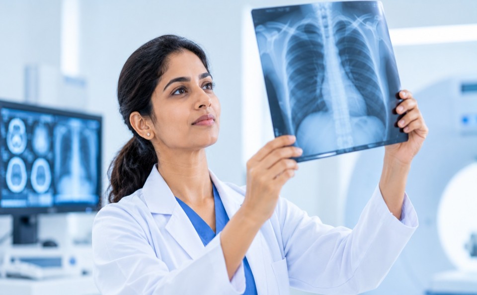
World Radiography Day: The Role of Medical Imaging in Early Diagnosis
Medical imaging plays an important role in modern healthcare by helping doctors understand what is happening inside the human body. From identifying bone fractures to detecting diseases affecting the brain, lungs, heart and other organs, imaging investigations support accurate diagnosis and timely medical treatment. World Radiography Day is observed on 8 November every year to recognise the discovery of X-rays and appreciate the contributions of radiographers and radiology professionals to healthcare.
On this occasion, let us understand how medical imaging supports early diagnosis, guides treatment decisions and improves patient care.
What Is Medical Imaging?
Medical imaging refers to techniques used to create pictures of structures inside the body. These images help doctors identify abnormalities, monitor diseases and plan appropriate treatment.
Medical imaging is commonly used for:
- Detecting fractures and bone abnormalities.
- Identifying infections and lung conditions.
- Examining the brain and nervous system.
- Evaluating abdominal organs.
- Detecting certain tumours and other abnormal growths.
- Monitoring the progress of diseases.
- Guiding certain medical procedures.
The type of imaging recommended depends on the patient’s symptoms, medical history and clinical examination.
Common Types of Medical Imaging
Different imaging techniques provide different types of information. Doctors select the most appropriate investigation depending on the condition being evaluated.
1. X-ray
X-rays use a small amount of ionizing radiation to produce images of structures inside the body.
They are commonly used to:
- Identify fractures and dislocations.
- Examine the chest and lungs.
- Assess certain bone and joint conditions.
- Detect some dental abnormalities.
Chest X-rays are frequently used to evaluate conditions such as pneumonia and other lung abnormalities.
2. Ultrasound
Ultrasound uses high-frequency sound waves to produce images of internal organs and soft tissues. It does not use ionising radiation.
It is commonly used for:
- Monitoring pregnancy.
- Examining abdominal organs.
- Evaluating the kidneys and urinary system.
- Assessing certain soft tissue abnormalities.
- Examining blood flow through Doppler ultrasound.
Ultrasound is particularly useful in obstetric and gynaecological examinations.
3. Computed Tomography (CT Scan)
A CT scan uses X-rays and computer processing to produce detailed cross-sectional images of the body.
CT scans may be recommended to:
- Evaluate head injuries and suspected brain bleeding.
- Assess chest and abdominal conditions.
- Identify certain tumours and infections.
- Examine complex fractures.
- Support emergency diagnosis.
CT imaging is particularly valuable in emergency situations where rapid assessment is required.
4. Magnetic Resonance Imaging (MRI)
MRI uses strong magnetic fields and radio waves to produce detailed images of organs and soft tissues. It does not use ionising radiation.
MRI is commonly used to examine:
- The brain and spinal cord.
- Muscles, ligaments and joints.
- Certain tumours and soft tissue conditions.
- Spinal abnormalities.
- Selected heart and blood vessel conditions.
Patients should inform the radiology team about implanted devices, metal objects or other relevant medical history before undergoing an MRI.
5. Mammography
Mammography is a specialised X-ray examination of the breast. It is an important screening and diagnostic tool for breast cancer.
It can help identify certain breast abnormalities, including suspicious lumps and changes that may not be felt during a physical examination.
The need and timing of mammography depend on age, symptoms and individual risk factors.
Why Is Medical Imaging Important for Early Diagnosis?
Many health conditions may not produce obvious symptoms during their early stages. Medical imaging can help doctors investigate suspected abnormalities and make informed clinical decisions.
Some important benefits include:
- Early identification: Helps detect certain abnormalities before symptoms become severe.
- Accurate diagnosis: Provides information about the location and nature of a suspected condition.
- Treatment planning: Helps doctors select suitable treatment approaches.
- Disease monitoring: Allows healthcare professionals to assess changes over time.
- Emergency assessment: Supports rapid evaluation of injuries and serious medical conditions.
- Procedure guidance: Helps guide selected biopsies, drainage procedures and other interventions.
Medical imaging is an important part of diagnosis, but it is not always required for every patient. Doctors recommend investigations based on clinical need.
Is Medical Imaging Safe?
Medical imaging is generally safe when performed for an appropriate medical reason using suitable protocols.
However, different imaging techniques have different safety considerations.
- X-rays and CT scans use ionising radiation. Healthcare professionals follow appropriate protocols to minimise unnecessary radiation exposure.
- Ultrasound does not use ionising radiation.
- MRI does not use ionising radiation but requires screening for certain implants and metal objects.
- Contrast agents used in some examinations may require additional precautions based on kidney function, allergies and medical history.
Pregnant patients or individuals who may be pregnant should inform their doctor and radiology team before undergoing imaging investigations.
Patients should always discuss their concerns with the healthcare professional performing or recommending the examination.
The Role of Radiologists and Radiographers
Medical imaging involves teamwork between different healthcare professionals.
Radiologists are doctors trained to interpret medical images and provide diagnostic reports. They work closely with treating doctors to support diagnosis and treatment planning.
Radiographers are trained professionals who perform imaging examinations, position patients correctly, operate imaging equipment and follow radiation safety procedures.
Together, they help ensure that imaging investigations are performed appropriately and provide useful diagnostic information.
When Should You Consult a Doctor for an Imaging Investigation?
Medical imaging should be guided by a doctor’s clinical assessment.
You may be advised to undergo an imaging investigation if you experience:
- Persistent or unexplained pain.
- Suspected fractures following an injury.
- Persistent cough or breathing difficulties.
- Severe headaches or neurological symptoms.
- Abdominal pain or swelling.
- Unusual breast changes or lumps.
- Symptoms requiring further investigation based on clinical examination.
In emergencies, such as suspected stroke, serious head injury or severe chest pain, seek immediate medical attention rather than delaying care to arrange an imaging appointment independently.
Choose the Right Investigation at the Right Time
Medical imaging has transformed modern healthcare by helping doctors examine internal structures, identify abnormalities and plan appropriate treatment.
However, the right investigation at the right time is essential. Unnecessary imaging should be avoided, and patients should follow the advice of qualified healthcare professionals.
Recognising the Contribution of Radiology Professionals
On World Radiography Day, we recognise the dedication and expertise of radiologists, radiographers and the entire radiology team who contribute to patient diagnosis and care. At East Point Hospital, medical imaging supports the evaluation and management of a wide range of health conditions. Patients can consult their treating doctors for guidance on appropriate diagnostic investigations.
Better imaging. Timely diagnosis. Improved patient care.